Exploring treatment options in cancer: tumor treatment strategies


Stages Of Non-Small Cell Lung Cancer
Doctors stage non-small cell lung cancer (NSCLC) according to how far it has progressed. Stages range from occult (or hidden) cancer to stage 4 cancer, in which multiple organs may be affected.
Several different staging systems for non-small cell lung cancer (NSCLC) are used worldwide, but the most widely used approach is the TNM system.
A staging system helps doctors determine the best treatment plan and helps individuals with cancer and their families understand the severity of the disease and the outlook.
This article will examine the TNM stages of NSCLC and the cancer traits and grouping for each stage.
The TNM system for NSCLC staging considers the size and location of the tumor, as well as what other parts of the body are involved:
In addition to the TNM classifications, doctors use six stages to further describe the cancer. Stages 1 through 4 are divided into substages, as shown in this table:
At the occult stage, the main cancerous tumor can't be found (TX). Cancer cells might be found in phlegm or other lung fluid but not in other tests. The cancer isn't thought to have traveled to the lymph nodes (NO) or other parts of the body (MO).
In stage 0, the tumor is contained to the top layer of the airways and not deep into other lung tissue (Tis). The cancer also hasn't spread to the lymph nodes (NO) or other parts of the body (MO).
Doctors divide stage 1 NSCLC into four further classifications:
Stage 1A1
The characteristics of stage 1A1 are as follows:
Stage 1A2
In stage 1A2, the tumor is between 1 cm and 2 cm across and hasn't affected the bronchi or penetrated the visceral pleura (T1b), which covers the surface of each lung. It also hasn't reached the lymph nodes (NO) or distant body parts (MO).
Stage 1A3
In stage 1A3, the tumor is between 2 cm and 3 cm across and hasn't migrated to the visceral pleura or the main branches of the bronchi (T1c). The cancer still hasn't reached the lymph nodes (NO) or other parts of the body (MO).
Stage 1B
In stage 1B, the tumor hasn't reached the lymph nodes (NO) or other body parts (MO) but has at least one of the following traits (T2a):
Stage 2 is broken down into two classifications:
Stage 2A
At this stage, the cancer is still contained in the lungs and hasn't spread to nearby lymph nodes (NO) or distant body parts (MO). However, the tumor has at least one of the following characteristics (T2b):
Stage 2B
In this stage, the tumor is no bigger than 3 cm across but hasn't migrated to the visceral pleura or the branches of the bronchi (T1). It has, however, spread to the lymph nodes in or around the lungs on the same side as the tumor (N1) but not distant parts of the body (MO).
Or, the tumor has at least one of the following traits (T2):
In stage 2B, the cancer may also have reached the lymph nodes in or near the lungs on the same side as the tumor (N1) but hasn't spread to distant body parts (MO).
Or, the tumor hasn't reached the nearby lymph nodes (NO) or the distant body parts (MO) but has at least one of these following characteristics (T3):
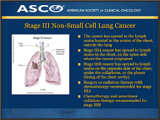
Stage three has three classifications:
Stage 3A
Stage 3 NSCLC is considered an advanced stage of the disease. However, it's treatable in many cases, with positive outcomes still possible. This stage covers a wide range of paths the cancer may be taking.
Path 1
In this case, the tumor is no more than 3 cm across, doesn't touch the main branches of the bronchi (T1), and hasn't reached the visceral pleura. It has spread to the lymph nodes on the same side as a main tumor (N2) but hasn't extended to distant body parts (MO).
Or, the tumor has at least one of the following traits (T2):
Path 2
In this case, the cancer has reached the lymph nodes near the main tumor (N2) but hasn't spread to distant body parts (MO).
Or, the tumor has at least one of the following features (T3):
Path 3
In this path of NSCLC, the cancer has spread to the nearby lymph nodes on the same side as the main tumor (N1) but hasn't reached other body parts (MO).
Or, the tumor has at least one of the following traits (T4):
The cancer may have spread to the lymph nodes in or near the lungs. If that has occurred, the lymph nodes are on the same side as the main tumor (NO or N1). The cancer hasn't spread to distant body parts (MO).
Stage 3A groupingStage 3B
This stage can also mean many different paths for the NSCLC.
Path 1
One possibility is that the cancer is no more than 3 cm across, hasn't entered the visceral pleura, and doesn't affect the main branches of the bronchi (T1).
However, the cancer has reached the lymph nodes close to the collarbone on either side of the body and may have also spread to the lymph nodes near the lungs on the side opposite the main tumor (N3). The cancer hasn't spread to distant parts of the body (MO).
Or, the tumor has at least one of the following traits (T2):
Path 2
In this scenario, the cancer has reached the lymph nodes close to the collarbone on either side of the body or has spread to the lymph nodes in or near the lungs on the opposite side from the main tumor (N3). This cancer hasn't reached distant parts of the body (MO).
Or, the tumor has at least one of the following traits (T3):
Path 3
In this case, the cancer has reached the lymph nodes near the carina or in the mediastinum on the side of the main tumor (N2) but hasn't traveled to distant parts of the body (MO).
Or, the tumor has at least one of the following characteristics (T4):
Path 4
In the path 4 scenario, the cancer has made its way to the lymph nodes surrounding the carina or in the mediastinum on the same side as the main tumor (N2). However, the cancer has not spread to distant body parts (MO).
Stage 3B groupingStage 3C
At this stage, the tumor has at least one of the following features:
The cancer has also spread to the lymph nodes close to the collarbone on either side of the body and may have reached lymph nodes on either side of the body from the main tumor (N3). The cancer hasn't reached distant body parts (MO).
Other characteristics of this stage
In this stage, the tumor may also have at least one of the following characteristics (T4):
The cancer may also have spread to the lymph nodes close to the collarbone on either side of the body and may have reached lymph nodes on either side of the body from the main tumor (N3). The cancer hasn't reached distant body parts (MO).
Stage 4A
At stage 4, the tumor may be any size, and the cancer may have extended into nearby tissue (any T). Likewise, it may not have reached nearby lymph nodes (any N).
For M1a grouping, at least one of the following traits must be present:
Or, the tumor may be any size, and the cancer may have extended into nearby tissue (any T). It may also involve any of the nearby lymph nodes (any N).
In addition, it has spread as one tumor outside of the chest cavity to a distant lymph node or an organ, such as the brain or liver (M1b).
Stage 4B
At stage 4, the tumor may be any size, and the cancer may have extended into nearby tissue (any T). Likewise, it may have reached nearby lymph nodes (any N).
The cancer has also spread as at least two tumors outside the chest cavity to distant lymph nodes and other organs (M1c).
As precise as these stages and groupings may seem, health experts acknowledge that classifying the state of a particular NSCLC is imperfect.
One 2017 study found that, as NSCLC progresses, staging accuracy declines. The authors recommend that an individual's healthcare team reassess the status of the cancer after every surgery or test to build a consensus about the next phase of treatment.
Clinicopathologic And Genomic Discoveries In Metastatic Lung Cancer
Despite the high incidence of brain metastasis after resection of non-small cell lung cancer (NSCLC), the National Comprehensive Cancer Network guidelines do not recommend central nervous system (CNS) surveillance. However, if patients with a high risk of brain metastasis after surgical resection can be identified, surveillance and prophylactic treatment with CNS-directed therapies may improve outcomes.
Memorial Sloan Kettering Cancer Center (MSK) researchers have made several recent discoveries about the clinicopathologic and genomic features predicting higher-risk lung cancer metastatic disease. These insights have revealed new treatment targets and are informing clinical trial risk stratification.
Stage 1 Lung Cancer: Prognostic Features Associated With a Higher Risk Of RecurrenceA team of 18 MSK experts, including thoracic surgeons and pathologists, has found that commonly reported features can be used to identify patients with stage 1 lung adenocarcinoma with a high risk of recurrence after complete resection.
Their retrospective study was the largest series globally to evaluate associations between clinicopathologic features and disease recurrence in this patient population. The paper was published in the February 2025 issue of the Journal of Thoracic and Cardiovascular Surgery. (1)
Led by thoracic surgeon David R. Jones, MD, Chief of the Thoracic Service and Co-Director of the Druckenmiller Center for Lung Cancer Research, the research team assessed results for 1,912 MSK patients who underwent R0 section for stage 1 lung adenocarcinoma from 2010 to 2020. (1)
The five-year cumulative incidence of recurrence was 12%. Among 250 patients who developed recurrence, 141 (56%) had distant and 109 (44%) had locoregional recurrence. (1)
Features independently related to a higher risk of recurrence were as follows: a higher maximum standardized uptake value of the primary tumor (hazard ratio (HR), 1.04); sublobar resection (HR, 2.04); higher lung cancer grade (HR, 5.32 for grade 2 and 7.93 for grade 3); lymphovascular invasion (HR, 1.70); visceral pleural invasion (HR, 1.54); and tumor size (HR, 1.30). Tumors with three to four high-risk features had a higher cumulative incidence of recurrence at five years of 30% versus 4% for tumors with fewer features. (1)
Key learning from this study included that tumor biological features associated with a poorer prognosis are more strongly associated with recurrence than tumor size. Moreover, aggressive features appeared to be additive. Improved risk stratification will help surgeons identify select patients who may benefit from inclusion in clinical trials of adjuvant therapies.
Moreover, the results of this retrospective study formed part of the basis for a recently opened global phase 3 clinical trial (NCT06564844) evaluating the effects of adjuvant immunotherapy and a bispecific antibody-drug-conjugate in patients with stage 1 NSCLC who are ctDNA-positive or have at least one high-risk pathological feature. Patients with EGFR or ALK alterations are excluded from the trial.
The retrospective study was funded by National Institutes of Health/National Cancer Institute grants. Access disclosures for Dr. Jones.
MSK researchers evaluated features associated with brain metastasis in a retrospective study of 2,660 patients who underwent complete resection of stage 1-3A lung adenocarcinoma between 2011 and 2020 at MSK.
This study was the largest to examine risk factors for brain metastasis after surgical resection of NSCLC and the only one to analyze the topic after resection for lung adenocarcinoma specifically. Led by Dr. Jones, the study was published in JTCVS Open in December 2024. (2)
The cumulative incidence of brain metastasis at 10 years was 9.8%, with a median time from surgery to metastasis of 21 months. Better performance status, lack of extracranial metastasis, stereotactic radiosurgery, and targeted therapy were associated with better survival. The median survival after brain metastasis was 18 months. (2)
Clinicopathologic features associated with a higher risk of brain metastasis were a higher maximum standardized uptake value on PET scan of the primary tumor, neoadjuvant therapy, lymphovascular invasion, and stage 3 disease. (2)
A subset of patients had next-generation sequencing performed on their primary tumor using MSK-IMPACT®. Neoadjuvant therapy, pathologic stage, and TP53 mutations were associated with the development of brain metastasis in this subgroup. (2)
The study was funded by grants from the National Institutes of Health/National Cancer Institute. Access disclosures for Dr. Jones.
MSK researchers recently discovered activation of both the TGF-beta and RAS signaling pathways is required for the development of lung cancer metastases.
Their study, published in Cell in September 2024, (3) represents years of work led by Joan Massagué, PhD, Chief Scientific Officer and Director of the Sloan Kettering Institute at MSK.
Dr. Massagué is known for his pioneering work, which revealed key insights into transforming growth factor-beta (TGF-beta) signaling processes that promote metastatic growth. For this study, Dr. Massagué and his team recruited collaborators from across the Sloan Kettering Institute and Memorial Hospital who contributed their multidisciplinary expertise.
The research team found that a transcription factor controlled by RAS plays a critical role in metastasis, known as the RAS response element binding protein 1 (RREB1). They discovered that RREB1 cooperates with a signaling complex called SMAD4 that is controlled by TGF-beta. (3)
Further, inhibiting RREB1 disabled metastatic processes in mouse models, suggesting it could be a potential new drug target for addressing metastatic lung cancer. (3) Read more.
Refer to the paper to review the extensive list of funding organizations that provided grants supporting this study, including the National Institutes of Health, various philanthropic foundations and programs, and the Fiona and Stanley Druckenmiller Center for Lung Cancer Research at MSK.
Patient-Derived Organoid Model Informs Immune-Priming Strategy for Individual PatientsLed by Dr. Jones, the MSK researchers created the first in vivo metastasis model to investigate the biology of human lung cancer metastases. The model represents a valuable resource for identifying genes related to brain metastasis. The paper was published in October 2024 in Cell Medicine Reports. (4)
For the model, they used lung adenocarcinoma patient-derived organoids with preserved biologic features of human metastases. Whole genome and RNA sequencing results showed that the model can be used to study clonality and tumor evolution and to identify biomarkers associated with metastasis to specific organ sites. (4)
The model helped the investigators determine the efficacy of treatments to suppress metastasis and identify mechanisms of drug resistance. For example, the model was highly useful in showing how KRASG12C- mutant lung adenocarcinoma responded to the targeted inhibitor sotorasib.(4)
Additionally, when cocultured with autologous peripheral blood mononuclear cells, the model demonstrated that it may help determine the optimal immune-priming strategy for individual patients with lung adenocarcinoma. (4)
The study was supported by grants from the National Cancer Institute, the National Institutes of Health, the Hamilton Family Foundation, and the Department of Defense. Access disclosures for Dr. Jones.
Read more about MSK's research into new treatment approaches for patients with lung cancer metastatic disease:
Refer a PatientCall our dedicated clinician access number at 646-677-7440 or click the link below, and one of our care advisors will assist you with your referral needs.


Comments
Post a Comment