Small Cell Lung Cancer (SCLC): Practice Essentials, Pathophysiology, Etiology

What Happens After A Lung CT Scan?
Michael S. Niederman, MD, is the lead academic and patient quality officer in the division of pulmonary and critical care medicine at Weill Cornell Medical Center in New York City; a professor of clinical medicine at Weill Cornell Medical College; and Lauder Family Professor in Pulmonary and Critical Care Medicine. He was previously the clinical director and associate chief in the division of pulmonary and critical care medicine at Weill Cornell Medical Center.
His focus is on respiratory infections, especially in critically ill patients, with a particular interest in disease pathogenisis, therapy, and ways to improve patient outcomes. His work related to respiratory tract infections includes mechanisms of airway colonization, the management of community- and hospital-acquired pneumonia, the role of guidelines for pneumonia, and the impact of antibiotic resistance on the management and outcomes of respiratory tract infections.
He obtained his medical degree from Boston University School of Medicine, then completed his training in internal medicine at Northwestern University School of Medicine, before undertaking a pulmonary and critical care fellowship at Yale University School of Medicine. Prior to joining Weill Cornell Medicine, he was a professor in the department of medicine at the State University of New York in Stony Brook and the chair of the department of medicine at Winthrop-University Hospital in Mineola, New York, for 16 years.
Dr. Niederman served as co-chair of the committees that created the American Thoracic Society's 1993 and 2001 guidelines for the treatment of community-acquired pneumonia and the 1996 and 2005 committees that wrote guidelines for the treatment of nosocomial pneumonia. He was a member of the American Thoracic Society/Infectious Diseases Society of America committee that published guidelines for community-acquired pneumonia in 2007. He was also the co-lead author of the 2017 guidelines on nosocomial pneumonia, written on behalf of the European Respiratory Society and the European Society of Intensive Care Medicine.
He has published over 400 peer-reviewed or review articles, and has lectured widely, both nationally and internationally. He was editor-in-chief of Clinical Pulmonary Medicine, is an associate editor of Critical Care and the European Respiratory Review, and serves on the editorial boards of Critical Care Medicine and Intensive Care Medicine. He has previously served on the editorial boards of the American Journal of Respiratory and Critical Care Medicine and Chest. For six years, he was a member of the Board of Regents of the American College of Chest Physicians, and in 2013, he was elected as a master of the American College of Physicians.
Presentation, Diagnosis And Assessment
Clinical PresentationLung cancer in general, and non-small cell lung cancer (NSCLC) in particular, is often asymptomatic in the early stages of the disease. When symptoms do occur in an early stage (e.G., endobronchial tumors), they include:
In addition to the above respiratory symptoms, advanced-stage NSCLC may present with more severe symptoms, including weight loss, fatigue and other systemic symptoms. The typical sites of metastasis for NSCLC include the brain, bones, liver, adrenal glands and the contralateral lung; metastasis related symptoms, such as pain or other symptoms (e.G., headaches or neurological symptoms for brain metastasis) may also occur. It is important to emphasize that these symptoms are not specific for lung cancer and are often mistaken for other conditions, which can lead to delays in diagnosis. At first diagnosis, the…
Clinical PresentationLung cancer in general, and non-small cell lung cancer (NSCLC) in particular, is often asymptomatic in the early stages of the disease. When symptoms do occur in an early stage (e.G., endobronchial tumors), they include:
In addition to the above respiratory symptoms, advanced-stage NSCLC may present with more severe symptoms, including weight loss, fatigue and other systemic symptoms. The typical sites of metastasis for NSCLC include the brain, bones, liver, adrenal glands and the contralateral lung; metastasis related symptoms, such as pain or other symptoms (e.G., headaches or neurological symptoms for brain metastasis) may also occur. It is important to emphasize that these symptoms are not specific for lung cancer and are often mistaken for other conditions, which can lead to delays in diagnosis. At first diagnosis, the typical patient with lung cancer, including NSCLC, is older (median age 71 years; <10% of cases are in people <55 years old), male (55%), and has a history of tobacco smoking (85-90%). Women with lung cancer are more likely to have been never-smokers than men.
DiagnosisEstablishing a definitive diagnosis of NSCLC requires several critical steps, outlined in Table 2-1. Any symptoms consistent with NSCLC should prompt further investigation.
A full medical history, including comorbidities, weight loss and physical examination, is required. Laboratory tests, including hematological, renal, hepatic and bone biochemistry tests, must also be conducted to assess the patient's overall health status and help select a treatment strategy.
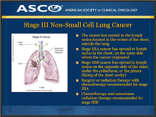
Imaging is crucial for both diagnosis and staging of NSCLC and can be performed utilizing several techniques, each with its own unique advantages and drawbacks. Traditionally, a simple chest X-ray (CXR) was used; however, while a CXR can still be used as the initial step in the diagnostic progress, the false-negative rate is unacceptably high (25%), and thus additional imaging must be utilized in the case of a negative result. A computed tomography (CT) scan provides additional information including the size, number and position of lung tumors; to assess lymph node involvement; and to locate potential extrathoracic metastases (Figure 2-1). As such, a CT scan is a useful modality for cancer staging. Lung cancer is often discovered incidentally on CT scans conducted for other purposes.
Patients without obvious extrathoracic metastasis may benefit from positron emission tomography (PET) scanning. This technique uses a radiolabeled glucose analog, fluorodeoxyglucose (FDG), to identify tissues with high metabolic activity (high glucose uptake), and can be used to risk-stratify the lesions identified on CT, stage mediastinal lymph nodes and identify distant metastases. A combination of FDG-PET and CT provides both anatomical and metabolic information and, thus, both improves diagnostic accuracy and prognostic value, and reduces the number of invasive procedures required for staging. A PET/CT radiograph is shown in Figure 2-2. Nevertheless, even with an FDG-PET/CT scan, false positives (e.G., due to infection, inflammation, or edema) and false negatives (e.G., when lesions are small or with other concurrent lung disease and diabetes) may occur.
While imaging techniques can detect and locate the presence of suspicious lesions that could be cancerous, a definitive diagnosis requires a morphological and histological analysis of putative tumors to determine if they are malignant. Before a biopsy is performed to sample the lesion, risk stratification of nodules identified on imaging should be performed using a validated model, such as the Mayo Clinic Solitary Pulmonary Nodule (SPN) Malignancy Risk Score. The SPN Malignancy Risk Score considers criteria such as age, history of smoking, prior extrathoracic cancer, nodule size, nodule location, and presence of spiculation (irregular, sharp, or jagged edges) on CT imaging. If a FDG-PET scan was performed, the level of FDG uptake can be added to the risk calculation. A score of <5% indicates low risk, that of 5-65% intermediate risk, and that of ≥65% high risk; a biopsy is recommended for intermediate and high risk nodules.
Many biopsy techniques exist that can be employed to obtain putative lung cancer samples. Endobronchial biopsy is a minimally invasive technique performed during bronchoscopy to sample visible tumors or lesions within the trachea or bronchi that can be accessed directly with the bronchoscope. Transbronchial lung biopsy (TBLB) uses a fluoroscopy-guided biopsy forceps, advanced into the peripheral lung via a bronchoscope, to sample lesions which are not directly visible. With TBLB, there is a small (1-5%) risk of complications such as bleeding or pneumothorax. Transbronchial needle aspiration (TBNA) uses a needle, again advanced through a bronchoscope, to penetrate central lung lesions or lymph nodes outside the bronchial wall and aspirate samples for biopsy. This is particularly useful for sampling mediastinal lymph nodes for staging purposes. Radial endobronchial ultrasound (EBUS)-guided biopsy uses a rotating ultrasound probe, passed through a bronchoscope, to locate peripheral lung nodules; once located, the lesion is marked and biopsied using tools like forceps or needles. Bronchial brushing is a method that uses a brush advanced through any working channel to scrape cells from visible lesions on the bronchial mucosa; this method is especially useful for cases where tissue biopsy is not necessary or feasible. Cytological specimens can also be obtained via bronchoalveolar lavage (BAL), which uses sterile saline introduced to a local lung segment through the bronchoscope for this purpose. Electromagnetic navigational bronchoscopy (ENB) uses a virtual 3D model of the patient's lungs (typically constructed using CT scan data) to guide a biopsy catheter through which biopsy tools can be advanced. Use of ENB is particularly suited for sampling of small, deep-seated lung nodules that are difficult to reach through traditional bronchoscopy but need precise navigation. In image-guided percutaneous fine-needle aspiration (FNA) or core-needle biopsy, a needle is inserted through the chest wall to sample a lung nodule under CT or ultrasound guidance. This method is used to access nodules near the lung surface, pleural-based lesions, or when bronchoscopy is not feasible. Core-needle biopsies are preferred to fine-needle aspirations when a larger amount of tissue is required for diagnosis. When large central airway tumors that require biopsy or debulking are involved, a rigid bronchoscope can be used to provide a wider working channel for biopsy. Other procedures, including EBUS-guided transbronchial needle aspiration, can be done through a rigid bronchoscope as well.
Once the radiographic data and biopsy samples have been obtained, a definitive diagnosis of cancer can be made, and the cancer can be typed. The first task is to distinguish cancerous from benign nodules. Benign nodules, such as granulomas or hamartomas, often have characteristic radiological features, including rounded, smooth, or polygonal contours and distinct calcification patterns (diffuse, central, laminar, or popcorn-like). By contrast, malignant nodules are characterized by features like lobulated or spiculated edges, pleural retraction and an eccentric calcification pattern (or no calcification). Suspicious nodules are definitively tested for malignancy by histological examination of biopsy specimens.
The second task is to type and grade the cancer. As described in Overview the most common types of NSCLC are adenocarcinoma, squamous cell carcinoma (SqCC) and lung carcinoma (LCLC). These can be distinguished from each other and from SCLC based on characteristic morphological features (see Overview) and the presence or absence of histological markers, as assessed by immunohistochemical staining of the biopsy sample:
In addition to providing the material to confirm the diagnosis and determine the histological type of the cancer, biopsies can also help in staging the cancer by determining whether the cancer has spread to the lymph nodes or other tissues. In addition, the material provided by the biopsy can be tested for the presence of biomarkers that can guide the choice of therapy.
A New Era In Cancer Care: Adventist Health Ukiah Valley Unveils New PET/CT Machine
Adventist Health Ukiah Valley recently unveiled its new PET/CT machine, which will offer faster results and peace of mind for patients and physicians. This addition is another crucial tool in the ongoing battle against cancer in our community and reinforces Adventist Health's commitment to providing high-quality, patient-centered, care close to home.
A positron emission tomography (PET) scan uses a radioactive substance called a tracer to look for disease in the body. PET/CT scans show detailed images and functions of internal organs for diagnosing conditions like cancer, heart disease and neurological disorders. This will be an important tool in diagnosing and treating cancers and will mean getting answers for our physicians and providing peace of mind for patients sooner.
"As a rural community, we've faced significant challenges in accessing timely cancer diagnostics. This is a monumental step forward for our cancer care and has truly been a long time coming. The arrival of a permanent PET/CT scanner here at Adventist Health Ukiah Valley significantly enhances our ability to diagnose and treat cancer more quickly and effectively. Early and accurate diagnosis is critical to improving outcomes, and this technology will have a direct and positive impact on the lives of our patients. This permanent installation removes many of the barriers our patients have faced for years. It brings cutting-edge technology close to home, eliminates the need for long-distance travel, shortens wait times, and allows us to deliver more timely, efficient, and personalized cancer care," explains Dr. Hengbing Wang, oncologist at Adventist Health Ukiah Valley's Cancer and Infusion Center.
The new PET/CT unit will offer greater access to care close to home. Previously, PET/CT scans were available only once a week, with the nearest alternative being more than an hour away in Santa Rosa and limited to one day each week. This will be the only permanent PET/CT unit available five days a week in Mendocino and Lake counties. This means quicker, more accurate diagnostics and better monitoring for those in the fight against cancer.
"This brings in a new era in cancer care for our community. Access to this important technology will improve outcomes for our patients and their families. Faster scanning will also improve efficiency and provide a more comfortable experience for our patients. We remain committed to delivering the latest advancements in diagnostic imaging, enhancing patient care, and improving access to care to our rural community," shares Jeff Mock, Operations Executive for Adventist Health Ukiah Valley.
Designed with the latest in imaging technology, the Canon PET/CT scanner provides exceptionally detailed images in about half the time of traditional scans. It delivers clearer images, which enable our physicians to make informed decisions regarding patient care and next steps. This advanced system also enhances patient comfort and safety with faster, lower-dose scans and improved diagnostic accuracy thanks to higher-resolution images.
A PET/CT scan can help with diagnosis/treatment monitoring for cancers such as: non-small cell lung cancer, colorectal cancer, esophageal cancer, head and neck cancers, lymphoma, ovarian cancer, myeloma, cervical cancer, breast cancer, thyroid cancer, and other types of cancers.
In addition to its value for cancer diagnosis and treatment, PET scans are also used for heart and neurological conditions to evaluate possible brain abnormalities, such as tumors, seizures and other central nervous system conditions.
To learn more about imaging services or to make an appointment, visit https://www.Adventisthealth.Org/ukiah-valley/services/imaging/


Comments
Post a Comment